In this Article
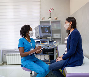
Medical diagnostic sonographers spend most of their day working one-on-one with patients. They capture images that doctors use to diagnose and treat illness, monitor conditions like pregnancy, and even assist in procedures such as surgery.
What Does a Sonographer Do?
“We use non-invasive technology and practices to aim sound waves at internal organs and blood vessels,” says Kate Scrivens, a sonographer in central Oregon. “The information we get by looking is used by a radiologist to understand what is going on with the patient.”
Since sonographers work closely with patients, they need to be caring and compassionate. Sometimes imaging is joyful—as in seeing a pregnant parent’s baby for the first—but some procedures are surrounded by anxiety. A good sonographer supports patients through both.
If you or someone you know has been pregnant, chances are you got your first glimpse of the baby in a sonograph. Sonographers create these images to determine whether a fetus is developing normally.
But a sonographer’s duties extend far beyond fetal scans. Sonographers also look for abnormalities such as kidney stones, cancers, and arterial blockages.
“We do just about everything,” Scrivens says. “We might start the day by looking at a fetus and a mom’s placenta, then check on another patient’s liver because of an abnormal blood test, then guide a biopsy. There are tons of different needs for sonographers.”
The work is physical, Scrivens says. Sonographers may need to help move or position patients and reach to move the ultrasound probe, or transducer, on the patient’s body to create the needed images.
Ultrasound works by emitting sound waves from the transducer that bounce off body fluid, tissue, and bone. The transducer receives these “echoes,” and the ultrasound translates the returning sound waves into a two-dimensional image. As the sonographer moves the transducer around, they make notes on the images they capture.
Roles and Responsibilities
“Sonographers are like the go-between for the patient and two doctors: the referring doctor and the radiologist,” Scrivens says. The referring doctor, who may be a primary care physician or a specialist, often sends notes on abnormal test results or indications that suggest a health problem. That information provides a starting point for a sonographer’s work.
As they go, sonographers adjust their imaging to make sure to capture all relevant images. “We are the radiologist’s eyes,” Scrivens says. “If we don’t document it, no one will see it.”
In some cases, especially ones that are complicated, unclear, or urgent, sonographers meet with the radiologist mid-exam. The radiologist will review the images captured up to that point and give guidance, which might include direction on whether or how to take additional images.
The radiologist may also step into the imaging room to explain findings, especially when results suggest something serious. Sonographers cannot deliver diagnoses; that responsibility falls to doctors.
A common misconception is that sonographers “just” take pictures. In reality, “we are highly trained professionals and absolutely know what we are looking at,” Scrivens says. They write summary reports after seeing each patient, they note what they suspect is going on with the patient—”and we’re expected to be right,” Scrivens says.
A Day in the Life of a Sonographer
A sonographer’s daily work depends partly on their workplace setting. “In most places, sonographers see a lot of variety,” Scrivens says. Sonographers can also work in a specialized setting, such as a vascular lab or lab for high-risk pregnancies.
In many cases, sonographers see patients back-to-back, followed by administrative time to write short summaries for each. A visit’s length depends on the type of imaging or procedure being performed.
“In a given day, I’ll do vascular studies, abdominal, guidance for procedures, babies, kids,” Scrivens says. “I end up doing a lot of everything.”
Most imaging is scheduled, which means sonographers must stay on schedule. “You have to balance between productivity and the quality doctors want,” Scrivens says. “You have to complete really complicated stuff in a certain time frame.”
Depending on where they work, sonographers may work weekends or be on call in case of an emergency.
Workplaces
“Your duties as a sonographer depend a lot on where you work,” Scrivens says. She has worked at a large trauma hospital, where she performed every type of ultrasound imaging. She’s also worked in a maternal-fetal medicine department, where she focused on fetal monitoring, high-risk pregnancies, and procedures such as amniocentesis.
Sonographers can work in a variety of settings.
- Hospitals:
- Most sonographers work in hospitals, either in a specialized lab or general imaging unit.
- Physicians’ offices:
- Some physicians’ offices, such as an OB/GYN clinic, may employ their own sonographers.
- Outpatient care centers:
- A small number of sonographers work in outpatient care, where patients receive treatment but do not need to stay overnight.
- Medical and diagnostic labs:
- Medical providers sometimes outsource imaging to medical and diagnostic labs, where sonographers often see a high volume of patients.
- Imaging equipment manufacturing and sales companies:
- Companies that design, manufacture, and sell ultrasound machinery employ sonographers to test equipment and even train customers.
Career Paths and Specialties
“Depending on where you work, you’ll either focus on one specialty such as vascular or OB/GYN, or you’ll do a huge variety,” Scrivens says. Specializing is possible but less common than being a generalist.
That said, “passing more board exams helps your job prospects,” Scrivens says. Earning credentials in a variety of specialties makes you more qualified for jobs that require broad and deep knowledge of diagnostic imaging.
Several sonographer specialties you may want to consider:
Abdomen
This is the most general specialty, and one of the essentials, Scrivens says. You’ll scan for things like fatty liver disease, cancers, obstruction of the bowels, and other conditions.
Obstetrics and gynecology
Pelvic ultrasounds can assess and monitor the health of a fetus as well as the patient’s placenta and cervix; they can also be used to detect conditions such as fibroids or cysts.
Cardiac
Imaging can identify heart conditions, such as damaged heart valves, and issues with heart function, such as decreased pumping strength.
Breast
Doctors may order breast ultrasounds as a follow-up to an inconclusive mammogram.
Vascular
Vascular ultrasounds are used to image veins and arteries to identify conditions such as blood clots.
Musculoskeletal
This type of imaging can be used throughout the body to help diagnose torn muscles or tendons, arthritis, or nerve issues.
What Makes a Successful Sonographer?
A desire to help others and an interest in technology could be considered the hallmarks of a good sonographer. But there are other traits and skills that are equally important:
- Compassion:
- Patients may feel scared or nervous, and they may be frustrated that you can’t tell them what you believe is wrong. You must stay empathetic and advocate to the radiologist and referring doctor on their behalf “so they get the best care possible,” Scrivens says.
- Physical fitness:
- Sonographers may need to stand for long periods, and they reach for much of the day. They also may lift, position, or move patients who need help, or move equipment from room to room.
- Poise under pressure:
- “You have to watch the time while you’re working so you’re not late, but also get the doctor what they need to diagnose the problem,” Scrivens says.
- Attention to detail:
- Your work must be precise, accurate, and thorough. “If we don’t image it, it’s missed,” she explains.
- Flexibility:
- The referring doctor will likely provide you with a list of suspected conditions, but you must be nimble enough to adjust your imaging based on what you see.
Education to Be a Sonographer
There are two primary paths to become a sonographer: an associate degree, which is most common, and a bachelor’s degree. For medical professionals who already have a health-related degree and are working in a clinical position, they have a third option of a certificate program, which takes one to two years.
If you are not already a medical professional, choosing between an associate or bachelor’s degree depends on several factors. You will need to weigh your finances, previous work and academic experience, career goals, and how quickly you want to start working.
Specialty Certifications
Although sonographers are not required to earn specialty certifications, most employers want new hires to be certified. “They’re a sign of being a professional and show you’re competent in that area,” Scrivens says.
In some cases, sonographers are hired under the agreement they will take and pass credentialing exams. Your employer may even pay for your exams.
Certifications may help your career by boosting your pay and opening doors to advancement. Scrivens says you’ll want to begin with the foundational credentials—in sonography principles and instrumentation, plus abdominal and OB/GYN. “Those are the essentials, and you can branch off from there,” she says.
Salary and Job Outlook
Sonography is a growing field with a good salary in return for two-year associate degree, if that’s the education route you choose. The U.S. Bureau of Labor Statistics (BLS) says sonography jobs will grow by 14.3% through 2032.
Much of this growth is due to baby boomers, who are not only aging but also living longer and requiring more care.
All in all, sonography is a promising career for people who are motivated and dedicated to helping others.

Written and reported by:
Catherine Ryan Gregory
Contributing Writer

With professional insight from:
Kate Scrivens
Diagnostic Medical Sonographer, Central Oregon Radiology